
Figure 4.
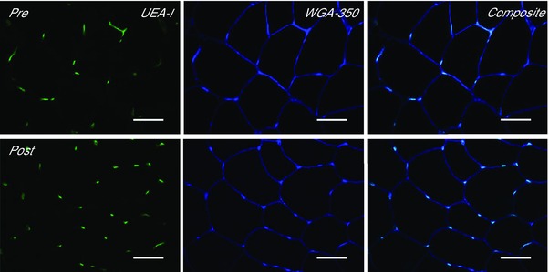
Effect of training on skeletal muscle capillarisation
Representative widefield microscopy images of skeletal muscle pre‐ (left) and post‐training (right). Skeletal muscle capillarisation was revealed using Ulex europaeus‐FITC conjugated lectin (UEA‐I, green), the skeletal muscle membrane was revealed using wheat germ agglutinin‐350 (WGA‐350, blue). Fibre type was revealed using anti‐myosin type I (image not shown). Composite image shows a combination of the UEA‐I and WGA‐350 images. Bar = 50 μm.