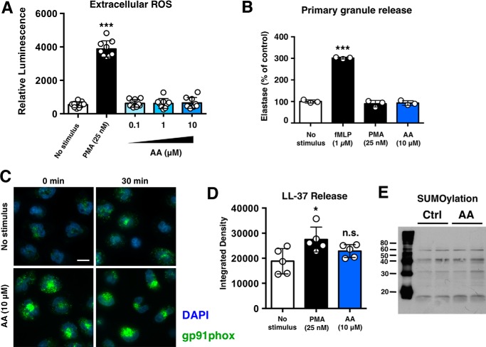
FIGURE 3.
Anacardic selectively stimulates specific neutrophil pro-inflammatory pathways. A, lucigenin-based assay to assess extracellular ROS levels following stimulation with anacardic acid (AA) or PMA (n = 9). B, assessment of neutrophil degranulation/elastase release using the colorimetric elastase substrate N-methoxysuccinyl-Ala-Ala-Pro-Val-p-nitroanilide. fMLP, N-formyl-Met-Leu-Phe. C, immunocytochemical analysis of gp91phox (green) in control and anacardic-acid treated neutrophils. Cell nuclei are stained with Hoerscht 33342 (blue). The scale bar represents 10 μm. D, dot-blot based assessment of LL-37 release from control, anacardic acid-treated, and PMA-treated neutrophils (n = 5). Anacardic acid does not stimulate neutrophil degranulation. E, anacardic acid does not inhibit SUMOylation in neutrophils (molecular masses shown are in kDa). Unless otherwise noted, data shown are expressed as mean values ± S.D. and are representative of at least three independent experiments performed in triplicate. Where applicable, the results were analyzed by one-way analysis of variance and post hoc Newman Keuls test. **, p < 0.01; ***, p < 0.001 versus control values. Ctrl, control.

