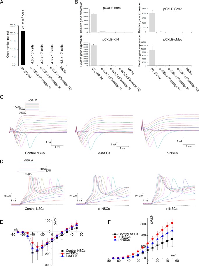
FIGURE 3.
Electrophysiological properties of e-iNSCs. A, the copy numbers of episomal vectors in e-iNSCs (passages 1, 5, and 10). Numbers indicate the number of estimated cell numbers in each cell. MEFs on day 5 post-transfection were used as a positive control. B, the episomal vector-mediated transgene expression in the established e-iNSCs (passages 1, 5, and 10) was examined by qPCR. The expression levels are normalized to those of MEFs. Error bars indicate standard derivation of duplicate values. C, representative voltage-clamp recordings in response to increasing voltage pulses, of which protocols are shown as the inset, from neurons derived from control NSCs, e-iNSCs, and r-iNSCs, respectively, after 14–16 days of differentiation. D, representative current-clamp recordings showing typical action potentials in response to increasing current pulses, of which protocols are shown as the inset, from neurons derived from control NSCs, e-iNSCs, and r-iNSCs, respectively. E and F, summary of the current-voltage (I-V) relationships for the voltage-gated Na+ (E) and K+ currents (F) in neurons derived from control NSCs (n = 3), e-iNSCs (n = 11), and r-iNSCs (n = 9).

