FIGURE 3.
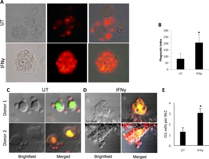
IFNγ enhances phagocytosis by NLCs. NLCs (n = 3 donors) were treated for 72 h with or without 10 ng/ml IFNγ and used in phagocytosis assays. A, representative microscopy images of untreated (UT, top panels) and IFNγ-treated (bottom panels) cells. Shown are bright-field (left panels), fluorescence (center panels), and merged (right panels) images. B, phagocytic index of untreated versus IFNγ-treated NLCs. C—E, NLCs (n = 5 donors) were treated as above and tested for phagocytosis of CLL cells as described under “Experimental Procedures.” Images show untreated (C) and IFNγ-treated NLCs (D). E, the average number of CLL cells ingested by NLCs. Error bars represent standard deviation. *, p ≤ 0.05.

