FIGURE 5.
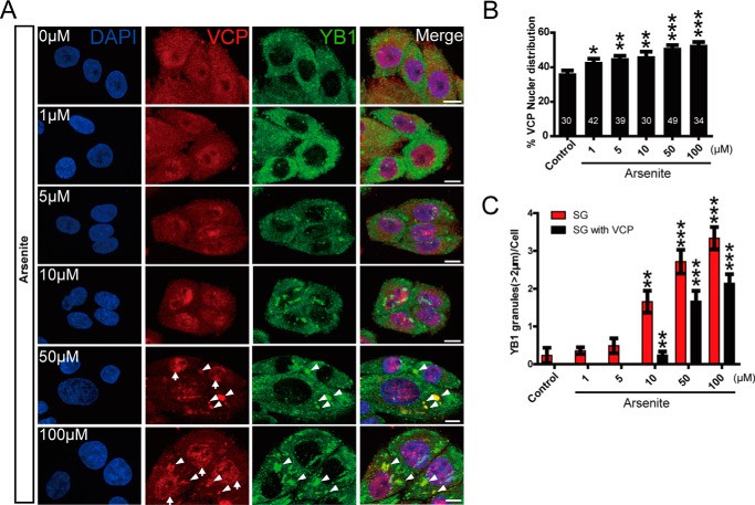
The accumulation of endogenous VCP in the nucleus and stress granules under stress condition. A, HeLa cells treated with increasing concentrations of sodium arsenite (2 h) were triple-labeled for VCP, DAPI, and stress granule marker YB-1 for confocal immunofluorescence microscopy analysis. Up arrows indicate nuclear VCP puncta, and arrowheads indicate VCP puncta in stress granules. Scale bar = 10 μm. B, the quantification of the fluorescence intensity of nuclear VCP, the number of cells (≥30) analyzed at each condition was labeled on each bar. C, quantification of the YB-1-positive stress granules (SG) (diameter ≥ 2 μm) per cell in cells exposed to sodium arsenite, with ≥30 cells analyzed at each condition.

