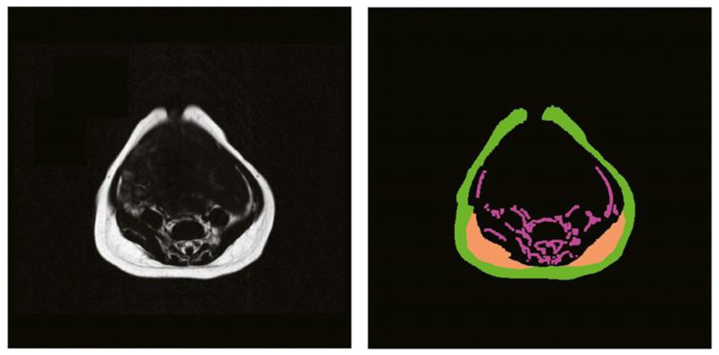
Figure 1.
Original abdominal water-suppressed MRI (left) and the results of the segmentation of abdominal adipose tissue compartments (right). Each compartment is filled with a different color as follows: green denotes superficial subcutaneous tissue, orange denotes the right and left deep subcutaneous tissue, and magenta denotes the internal adipose tissue.

