. Author manuscript; available in PMC: 2016 Jul 5.
Published in final edited form as: ACS Nano. 2016 Feb 1;10(3):3214–3221. doi: 10.1021/acsnano.5b05211

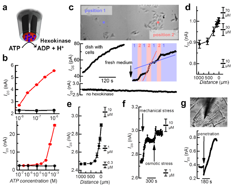
Figure 4. PPy nano-FET probe detects pH changes caused by hexokinase to perform highly localized ATP measurements for cell analysis. Immobilizing hexokinase to the tip of the probe creates a highly sensitive biosensor (a). Calibration curves are shown for two individual sensors, black-dot traces show the control experiment of a nano-FET not modified with hexokinase (b). Real-time ATP measurements over a nonpopulated spot and a group of melanoma cells demonstrate the high rate of ATP release from the cells (c). ATP gradients released from an isolated melanoma cell are measured by vertical approach of the sensor (d). Localized ATP measurements are also performed on a single cardiomyocyte (e–g). The cardiomyocyte produces a vertical gradient from its proximate microenvironment into the bulk solution (e). Additional ATP release is induced by touching the cell with the sensor and adding water to the solution to create osmotic stress (f). The probe allows for penetration into the cardiomyocyte (g).

Figure 4