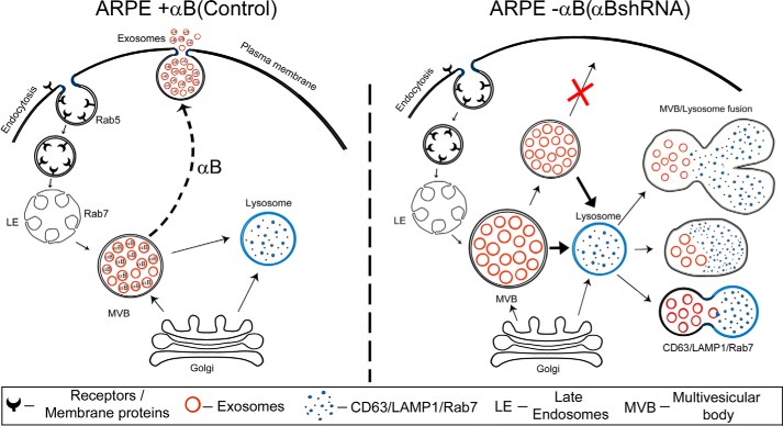
FIGURE 10.
Schematic representation of exosome biogenesis in presence and absence of αB expression in ARPE cells. The left schematic shows facilitation of exosome secretion by αB in native ARPE cells (αB on the dotted arrow pointing to the plasma membrane). Note that we have shown αB inside the exosomes based on previously published work (22). It remains to be established whether all the exosomes contain αB; therefore, we have shown about 20% of the exosomes without αB. The right schematic shows enhancement of the endolysosomal compartment (fused vesicles) in the absence of αB in ARPE-αB shRNA cells. Note that this enhancement is consistent with the increase in the LAMP1 (Fig. 8) and Rab7 labeling in this compartment (Fig. 9).

