
FIGURE 2.
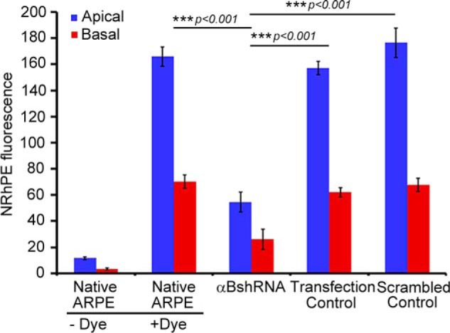
Inhibition of exosome secretion from apical and basal surfaces in αB silenced ARPE cells. Confluent monolayers of ARPE19 cells grown on transwell inserts were used to study the apical and basal exosome production using the NRhPE dye, a fluorescent lipid that is internalized by endocytosis and labels exosomes/MVBs (32). The cells were incubated with the NRhPE dye in serum-free medium, washed, and then incubated in the dye-free medium. Exosome preparations obtained from apical and basal medium were used to measure the total fluorescence as an indicator of exosome secretion into the medium (see under “Experimental Procedures”). The ARPE cells (αBshRNA), where αB expression is inhibited, show appreciable decrease in total fluorescence from both apical as well as basal surfaces indicating inhibition of exosome secretion from the apical as well as basal surfaces. αBshRNA = ARPE cell clone where αB expression was inhibited (Fig. 1A, αB shRNA, lane 2); Transfection control = αB shRNA transfected ARPE where there was no inhibition of the expression of αB (Fig. 1A, Transfection control); Scrambled control = ARPE cells transfected with a scrambled αB shRNA sequence that does not inhibit αB expression (Fig. 1A, scrambled control).