
FIGURE 3.
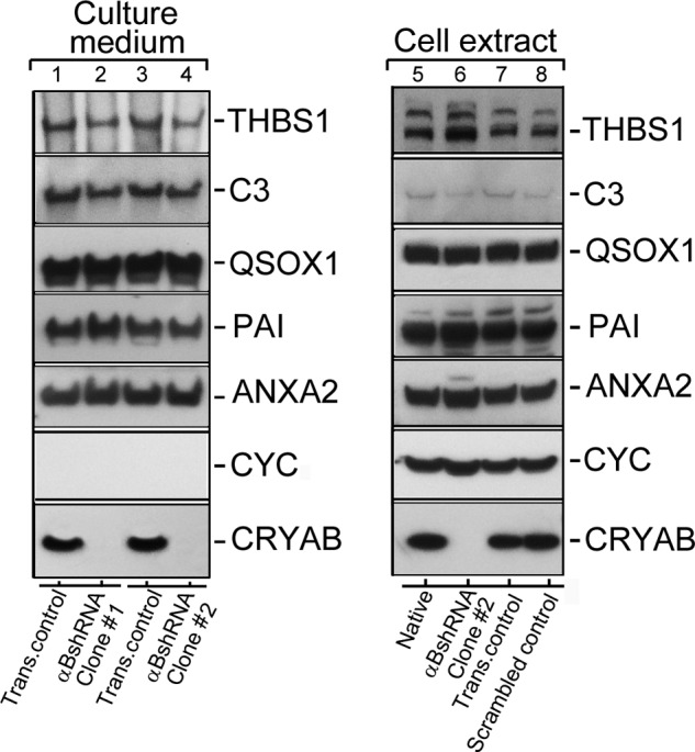
Conventional protein secretion in αB-silenced ARPE cells. Immunoblots of known extracellular proteins of the ARPE19 cells (in the culture medium and cellular extracts) are shown. 40 μg of protein were run on each lane on an SDS-polyacrylamide gel (gradient 4–12%; Life Technologies, Inc.), transferred to a membrane, and probed with following polyclonal antibodies (left panel shows the Culture medium, and the right panel shows total Cell extract immunoblots, respectively): anti-THBS1 (thrombospondin 1); anti-C3 (complement component 3); anti-QSOX1 (quiescin Q6 sulfhydryl oxidase 1); anti-PAI (serpin peptidase inhibitor, clade E (nexin, plasminogen activator inhibitor type 1), member 1); anti-ANXA2 (annexin II); anti-CYC (cytochrome c); and anti-αB (αB-crystallin, CRYAB). Two different clones of ARPE19 cells, permanently transfected with αB shRNA plasmids, that do express αB (lanes 2 and 4) were analyzed (Culture medium; left panel). All five proteins that were expected to be in the medium (as per conventional protein secretion) were detected in transfection control (Trans. control, lanes 1 and 3). Lanes 2 and 4 show absence of αB in the medium of ARPE αB shRNA clones #1 and #2 respectively. Note that cytochrome c (CYC) is not detected in the culture medium of all four cultures indicating that there is no cell death. The right panel shows immunoblots of the cell extracts from the Native un-transfected cells (lane 5), αBshRNA clone #2 (lane 6, αB-crystallin is silenced), transfection control (lane 7), and Scrambled shRNA control (lane 8), where αB-crystallin is not silenced. Based on these data, conventional protein secretion is not directly impacted by the absence of αB expression. In this immunoblot only αB shRNA clone #2 was used (lane 6).