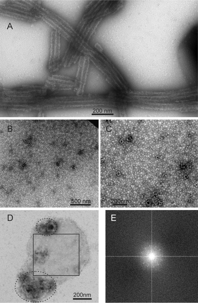
FIGURE 1.
TEM images of negatively stained HIV-1 capsid protein assemblies. A, WT-CA tubes. B and C, R18L-CA spheres. D, planar R18L-CA sheet. Non-planar material within ellipses may result from disruption or dissolution of R18L-CA sheets during the TEM grid preparation process. E, Fourier transform of the region of panel D enclosed by a square. The pattern of spots with 6-fold symmetry indicates two-dimensional crystallinity of R18L-CA sheets.

