FIGURE 2.

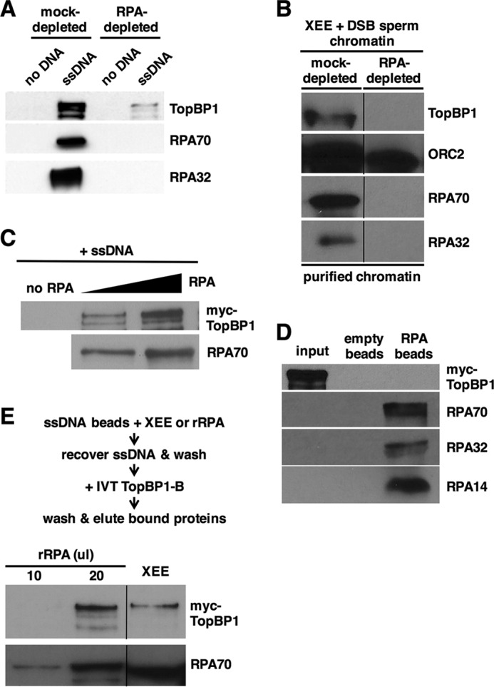
TopBP1 binds RPA-ssDNA, and not ssDNA alone or free RPA. A, same as Fig. 1A except Xenopus egg extract was either mock-depleted or depleted of RPA using anti-RPA antibodies as indicated. Data shown are representative of a minimum of two independent biological replicates. B, Xenopus egg extract (XEE) was either mock-depleted or depleted of RPA using anti-RPA antibodies and then mixed with sperm chromatin and EcoRI to induce DSBs into the chromatin. After a 1-h incubation the chromatin was isolated and probed for the indicated proteins by Western blotting. TopBP1 and the RPA proteins were detected as in Fig. 1A, and Orc2, which serves as a chromatin loading control, was detected with antibodies raised against Xenopus Orc2. The data shown are from different regions of the same gel, and the images were then spliced together, as indicated by the black line, so as to remove irrelevant material. Data shown are representative of a minimum of two independent biological replicates. C, IVT myc-tagged TopBP1 was mixed with varying amounts of purified, recombinant RPA, and biotin-coupled ssDNA (1.5 kb). The ssDNAs were isolated using streptavidin magnetic beads, washed, and eluted, and bound proteins were detected by Western blot. TopBP1 was detected using mAb 9E10, which recognizes the myc tag, and RPA was detected using antibodies against the Xenopus RPA trimer. Data shown are representative of a minimum of two independent biological replicates. D, purified, recombinant RPA trimer was optionally coupled to nickel-nitrilotriacetic acid agarose beads by virtue of a 6-histidine tag on the 70-kDa subunit. Beads were mixed with IVT myc-tagged TopBP1 and washed, and bound proteins were detected by Western blot. TopBP1 and the individual RPA subunits were detected as in C. Input refers to 5% of the initial reaction volume. Data shown are representative of a minimum of two independent biological replicates. E, biotin-linked ssDNA was mixed with either egg extract or differing amounts of purified recombinant RPA (rRPA). The ssDNAs were isolated using streptavidin magnetic beads, washed, and then incubated with IVT myc-tagged TopBP1. The beads were isolated again and washed, and bound proteins were detected as in C. The data shown are from different regions of the same gel, and the images were then spliced together, as indicated by the black line, so as to remove irrelevant material. Data shown are representative of a minimum of two independent biological replicates.
