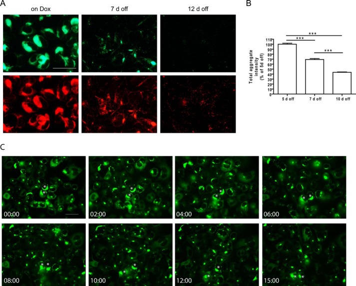
FIGURE 4.
Clearance of tau aggregates in clone 4.1 cells following suppression of soluble tau expression. A, Triton-insoluble tau aggregates labeled by GFP signals (top panels) and PHF-1 immunostaining (bottom panels) in clone 4.1 cells maintained on Dox or at 7 or 12 days off Dox. Scale bar, 50 μm. B, whole coverslip quantifications for the total intensity of Triton-insoluble GFP-positive tau aggregates in clone 4.1 cells at 5, 7, and 10 days off Dox. Cells were plated onto the coverslips at 5 days off Dox and no further passaging was performed thereafter. Three independent sets of experiments were performed, with the total aggregate intensity measurements normalized to that of 5 days off Dox within each set of experiment. Data are shown as mean ± S.E. One-way ANOVA was performed followed by Tukey's post hoc test for all pairwise comparisons. ***, p < 0.001. C, selected snapshots from live imaging of clone 4.1 cells from 4 days off Dox to 5 days off Dox. Stars mark examples of large and elongated aggregates getting consolidated into smaller round inclusions. See supplemental Movie S3 for the complete time-lapse video. Relative timing of the snapshots was presented as hh:mm. Scale bar, 50 μm.

