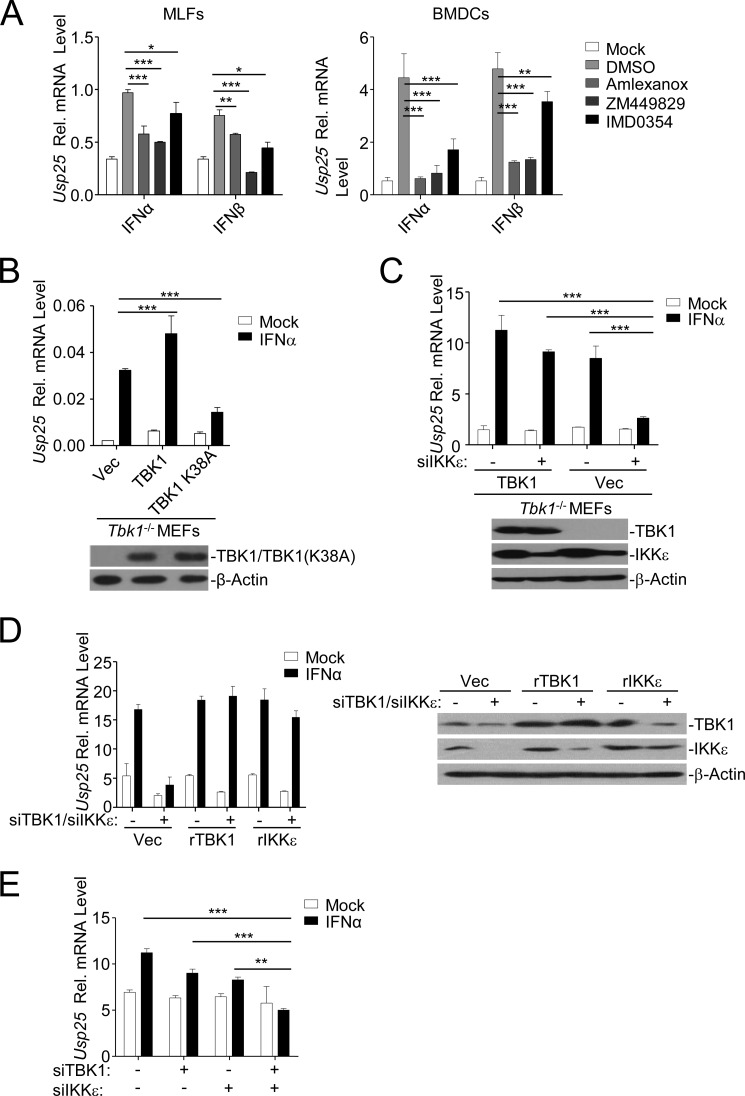
FIGURE 4.
TBK1 and IKKϵ function redundantly for type I IFN-triggered induction of Usp25. A, MLFs (left graph) or BMDCs (right graph) were left untreated or treated with IFNα (20 units/ml) or IFNβ (100 ng/ml) in the presence or absence of various kinase inhibitors for 12 h followed by qPCR analysis. B, Tbk1−/− MEFs were reconstituted with Vec, TBK1, or TBK1(K38A) through lentivirus-mediated gene transfer. The reconstituted TBK1 or TBK1(K38A) in the cells was examined by immunoblotting analysis (lower panels). Cells were left untreated or treated with IFNα (20 units/ml) for 12 h followed by qPCR analysis (upper graph). C, Tbk1−/− MEFs were reconstituted with Vec or TBK1 through lentivirus-mediated gene transfer. Cells were further transfected with control or siRNA targeting IKKϵ followed by immunoblotting analysis (lower panels) or qPCR analysis after IFNα treatment (20 units/ml) (upper graph). D, wild-type MEFs were stably transfected with Vec, rTBK1, or rIKKϵ followed by transfection of control or siRNAs targeting endogenous TBK1 and IKKϵ. Cells were subjected to immunoblotting analysis (right panels) or qPCR analysis after IFNα treatment (20 units/ml) (left graph). E, wild-type BMDCs were transfected control, siTBK1, or siIKKϵ. Twenty-four hours later, cells were stimulated with IFNα (20 units/ml) for 8 h followed by qPCR analysis. Data shown are representatives of at least three independent experiments. *, p < 0.05; **, p < 0.01; ***, p < 0.001. Error bars represent S.D. Rel., relative.

