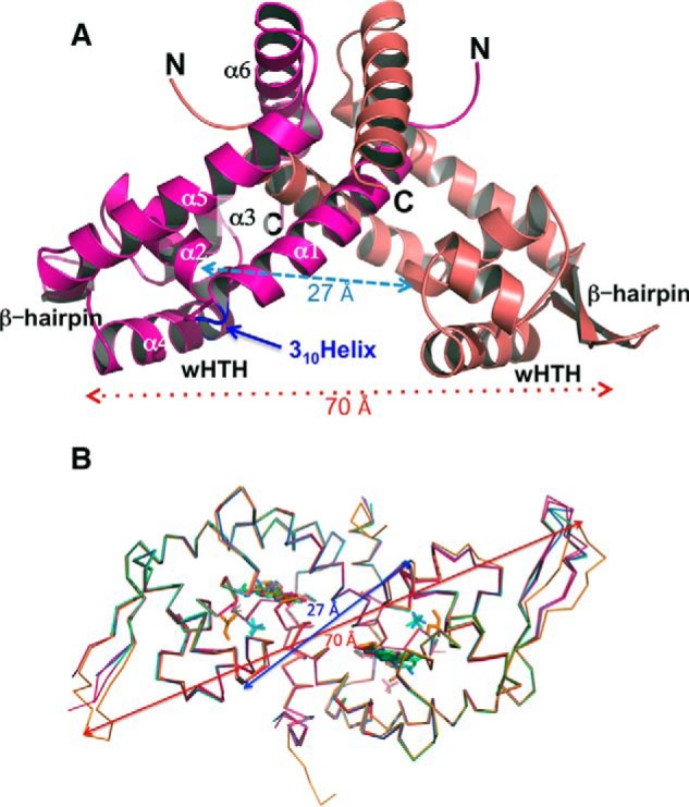
FIGURE 3.

Overall structures of AcHcaR dimers. A, ribbon diagrams of the AcHcaR dimer in stick representation. N and C termini and wHTH motifs are labeled. B, comparison of main chain atom conformation in AcHcaR structures. The main chain structure of apo-AcHcaR (red) is compared with AcHcaR complexes with ligands (AcHcaR·ferulic acid (orange), HcaR·DHBA (pink), HcaR·vanillin (blue), and HcaR·p-coumaric acid (green)). Ligands are shown as sticks using the same color scheme. In addition to aromatic compounds, glycerol and phosphate anions are found in some protein structures. Structures were superimposed with Coot (50). The arrows show distances between Cβ atoms of His91A-His91B (red) and Gln71A-Gln71B (blue). These residues are part of the wHTH DNA-binding motif.
