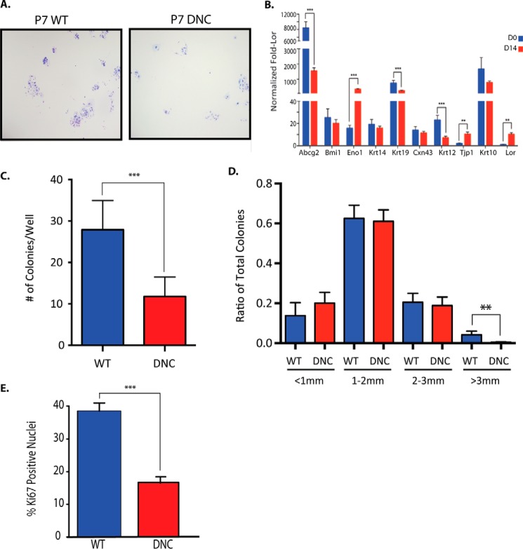
FIGURE 3.
Corneal epithelial progenitor cells are decreased in K14-DN-Clim mice at P7. A, Giemsa stained representative images of colony forming assays from P7 WT and DN-CLIM (DNC) primary corneal epithelial colonies (4×). B, comparison of expression levels across different marker genes at Day 0 (D0) and Day 14 (D14), normalized to loricrin (Lor) expression levels. All data were also normalized to 18S RNA. C, quantification of the number of colonies/well for WT and DN-CLIM corneal epithelial cells (n = 7 WT and 7 DN-CLIM mice). D, the ratio of colonies by size from P7 WT and DN-CLIM cells (n = 7 WT and 7 DN-CLIM mice). E, percentage of ki67-positive nuclei in WT and DN-CLIM corneal epithelial colonies. A t test was used to determine significance: **, p = 0.01; ***, p = 0.001. Error bars represent S.E.

