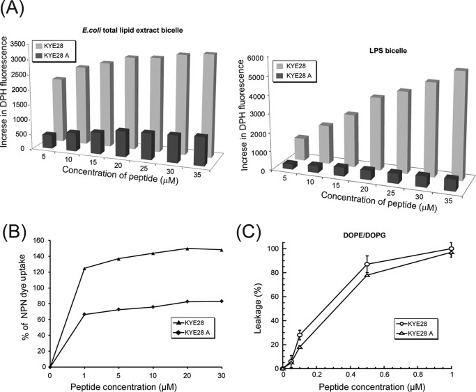
FIGURE 4.
Membrane permeabilization effects of KYE28 and KYE28A. A, bar plots showing the increase in DPH fluorescence as a function of peptide concentration upon addition of peptide to E. coli total lipid extract bicelle (left) and LPS bicelle (right) as a measure of bicelle disruption. KYE28 is seen to cause a pronounced concentration-dependent increase in DPH fluorescence in both E. coli total lipid extract bicelles and LPS bicelles, much larger than those caused by KYE28A. B, plot showing percentage of NPN dye uptake by X. vesicatoria cells upon addition of KYE28 and KYE28A. More than 2-fold higher uptake is seen for KYE28 as compared with KYE28A. C, percentage of dye leakage from DOPE/DOPG (75/25 mol/mol) vesicles as a function of peptide concentration. Both KYE28 and KYE28A induced comparable dye leakage from vesicles, KYE28 producing slightly greater effects.

