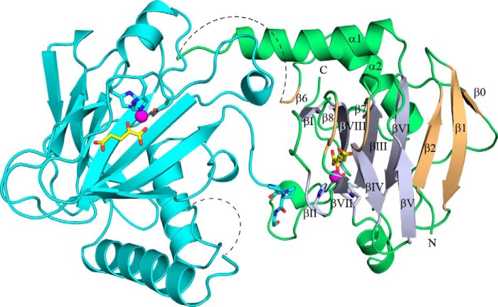
FIGURE 5.
Structure of the Co(II)-BaP4H-PPG monomer (green) showing the C terminus of a symmetry related molecule (cyan) interacting with the active site of chain A (green). The β-strands of the DSBH-fold (light blue) are labeled βI-βVIII. Other β strands (light orange) are labeled β0, β1, β2, β6, β7, and β8. Missing residues in the substrate binding loop region are indicated by a dashed line. The metal binding residues, His127, Asp129, and His193, αKG (yellow), and the fused (P-P-G) peptide are shown as sticks (oxygen, red; nitrogen, blue). Cobalt is shown as a magenta sphere.

