
FIGURE 8.
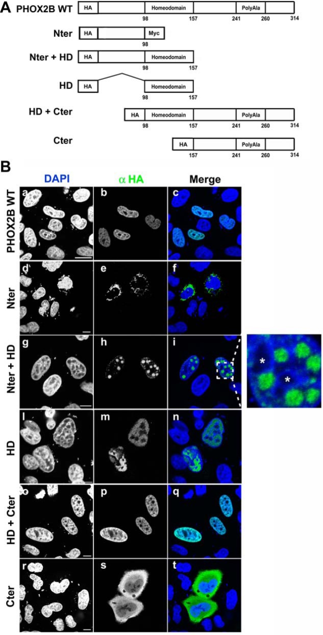
Contribution of the N- and C-terminal domains to nuclear import of PHOX2B. A, schematic representation of wild-type PHOX2B protein and its truncated constructs. All constructs were fused N-terminally to an HA epitope tag. Numbers correspond to the amino acids residues of PHOX2B. B, representative immunofluorescence images of the localization of HA-PHOX2B truncated fusion proteins. HeLa cells were transfected with the HA-tagged proteins and analyzed 48 h after transfection by means of immunofluorescence using anti-HA antibody (b, e, h, m, p, and s); the nuclei were visualized using DAPI (a, d, g, l, o, and r) and merged with the proteins detected by the anti-HA antibody (c, f, i, n, q, and t). On the right of i, an enlarged view of the indicated area is shown; the asterisks indicate the nucleoli.