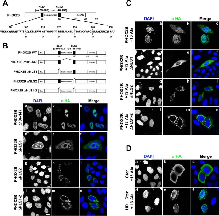
FIGURE 9.
Identification of PHOX2B NLSs and effects of the polyalanine-expanded tract on PHOX2B nuclear import. A, schematic representation of the PHOX2B protein showing the sequence of the homeodomain and the two putative NLSs (underlined). B, top, schematic representation of PHOX2B carrying deletions of the proximal NLS (ΔNLS1), distal NLS (ΔNLS2), both (ΔNLS1-2), or the entire HD except for the NLSs (Δ106–147). Black boxes, NLSs. Bottom, representative immunofluorescence images of the subcellular localization of HA-tagged PHOX2B deletion proteins in transfected HeLa cells stained by anti-HA antibody (b, e, h, and m); the nuclei were visualized using DAPI (a, d, g, and l) and merged with the HA-PHOX2B deleted proteins detected by anti-HA antibody (c, f, i, and n). Scale bars, 10 μm. C, representative immunofluorescence images of the subcellular localization of HA-tagged PHOX2B carrying +13 alanine expansions (PHOX2B +13Ala) and the expanded deletion proteins (PHOX2B +13Ala ΔNLS1, PHOX2B +13Ala ΔNLS2, and PHOX2B +13Ala ΔNLS1-2) in transfected HeLa cells stained by anti-HA antibody (b, e, h, and m). The nuclei were visualized using DAPI (a, d, g, and l) and merged with the HA-PHOX2B deleted proteins detected by anti-HA antibody (c, f, i, and n). Scale bars, 10 μm. D, representative immunofluorescence images of the localization of the expanded HA-PHOX2B truncated fusion proteins. HeLa cells were transfected with the HA-tagged proteins and analyzed 48 h after transfection by means of immunofluorescence using anti HA antibody (b and e); the nuclei were visualized using DAPI (a and d) and merged with the proteins detected by the anti-HA antibody (c and f). Scale bars, 10 μm.

