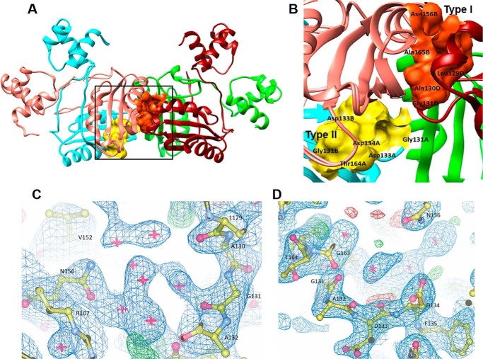
FIGURE 2.
Ligand binding sites of MtbAldR. A, type I binding site at the interdimer interface and type II binding site at the intradimer interface of MtbAldR are shown in orange and yellow, respectively. The region marked by the box has been expanded and shown in B, which shows details of the residues that make up the respective binding sites. Gly131 is common to both sites. 2|Fo| − |Fc| electron density maps contoured at 1.0σ at the type I binding site (C) and the type II binding site (D) are also shown.

