FIGURE 5.
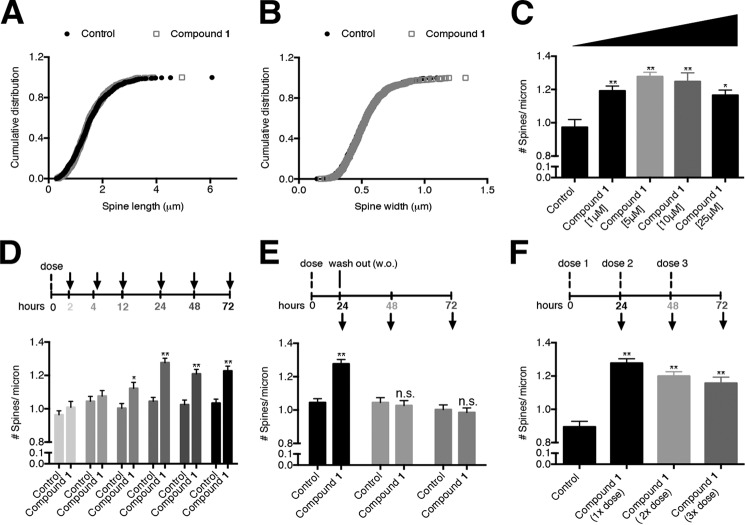
Examination of the spinogenic properties of BAM1-EG6 observed in rat primary hippocampal neurons. Cumulative distribution of spine length (A) or width (B) of control cells versus cells treated with compound BAM1-EG6 (1 μm). C, concentration-dependent effects of neurons dosed for 24 h with 1–25 μm BAM1-EG6 on spine density. D, kinetics of spine density increase in cells exposed to BAM1-EG6 compared with vehicle control (0.1% DMSO). Neurons were dosed and then fixed at 2, 4, 12, 24, 48, and 72 h. E, effects of removal of BAM1-EG6 on dendritic spine number after treatment of cells for 24 h. After 24 h, BAM1-EG6 was washed out (w.o.) and spine changes were monitored for an additional 24 and 48 h (48 and 72 h total time). The dendritic spine density 24 h after removal of BAM1-EG6 is indistinguishable from control cells. F, effect on spine density increases of adding additional doses of BAM1-EG6 every 24 h over a total incubation time of 72 h. Neurons were dosed at 24 h (1×), 48 h (2×), and 72 h (3×) with no observable additional increase of dendritic spine density compared with the 1× dose. Data are expressed as mean values ± S.E., n ≥ 54. **, p ≤ 0.0001; n.s., not significant, as determined by unpaired t test compared with control. Arrows denote time points when aliquots of cells were fixed and analyzed.

