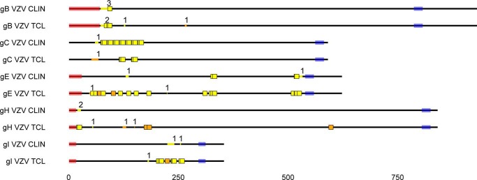
FIGURE 2.
O-Glycosites found in VZV derived from infected fibroblasts (TCL) and a clinical sample. VZV (strain Dumas) gB, gC, gE, gH, and gI protein sequences are shown as black lines, drawn to scale. Predicted signal peptides and transmembrane regions are shaded in pink and blue, respectively. Unambiguous O-glycosylation sites are shown as colored squares, whereas ambiguous sites are marked as colored lines within the protein backbone, where the number above indicates the number of glycosites. Trypsin and unique chymotrypsin digestion-derived glycosites are marked in yellow and orange, respectively. All identical potentially glycosylated VZV tandem repeats are shown occupied. Reference VZV sequences were used because of unavailable annotation of investigated isolate sequences. Glycoprotein M is not shown, because it was only found glycosylated in the infected cell lysate. CLIN, clinical sample; TCL, total infected cell lysate.

