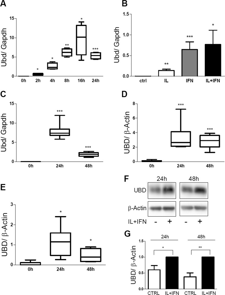
FIGURE 3.
IL-1β + IFN-γ induce UBD expression in rodent and human beta cells. The expression of UBD was assessed by RT-PCR (A–E) and by Western blot (F and G) in INS-1E cells (A and B), FACS-purified rat beta cells (C), human islet cells (D), and the human EndoC-βH1 cells (E–G), and normalized by the housekeeping gene Gapdh (A–C) or β-actin (D–G). Cells were left untreated or treated with IL-1β + IFN-γ (IL+IFN) at different times, as indicated (A and C–G) or left untreated or treated with IL-1β (white bar), IFN-γ (gray bar), and IL-1β + IFN-γ (IL+IFN; black bar) for 24 h (B). Results are represented as a box plot indicating lower quartile, median, and higher quartile, with whiskers representing the range of the remaining data points (A and C–E). One representative Western blot for UBD (F) and optical density analysis of 6–7 independent experiments are shown (G). Data were normalized against the highest value (considered as 1) in each independent experiment (G). *, p < 0.05; **, p < 0.01; ***, p < 0.001 versus 0 h or control (ctrl); paired Student's t test. Data shown are mean ± S.E. of 3–7 independent experiments.

