FIGURE 4.
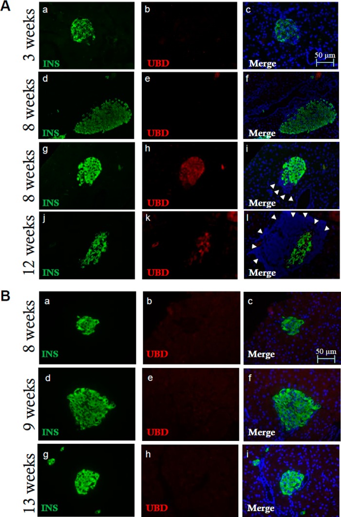
Increased expression of UBD in inflamed islets from diabetes-prone NOD mice. Pancreatic sections from 3-, 8-, and-12 week-old NOD mice were stained for insulin (panels a, d, g, and j, green), UBD (panels b, e, h, and k, red), and Hoechst for nuclear staining (panels c, f, i, and l, blue). Infiltrated lymphocytes, a sign of insulitis, are indicated by white arrowheads (panels i and l). Insulitis is present at 8 and 12 weeks of age but not at 3 weeks. The immunofluorescence analysis shows UBD expression in insulin-positive cells in NOD mice at 8 and 12 weeks (panels i and l, yellow) but not at 3 weeks (panels b and c) or at 8 weeks when insulitis is not present (panels e and f). The images shown are representative of 16 ± 3 islet sections from 3 to 4 different mice per age (A). Pancreatic sections from 8-, 9-, and 13-week-old NOD-SCID mice were stained with antibodies specific for insulin (panels a, d, and g, green), UBD (panels b, e, and h, red), and with Hoechst for nuclear staining (panels c, f, and i, blue). Immunofluorescence analysis indicates absence of UBD expression in insulin-positive cells in NOD-SCID mice (panels c, f, and i, merge). The images shown are representative of 12 ± 3 islet sections from 1 to 3 mice per age (B). Bars, 50 μm.

