FIGURE 2.

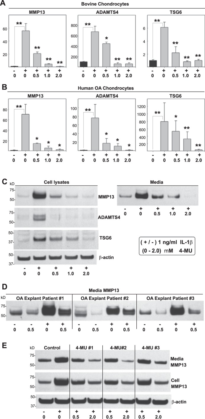
Effect of 4-MU on chondrocytes stimulated with IL-1β. High density monolayer cultures of bovine chondrocytes (A), human OA chondrocytes (B, C, and E), or explant cultures of human OA cartilage tissue cores (D) were treated without or with 1.0 ng/ml IL-1β (−/+) and co-treated with 0 to 2.0 mm concentrations of 4-MU as shown. After 24 h, the chondrocytes were lysed and analyzed by real time qRT-PCR (A and B). The fold changes (y axes) in mRNA copy number for MMP13, ADAMTS4, and TSG6 were compared with control (black bars) and quantified using the ΔΔCt approach with normalization to GAPDH. For statistical analysis, a two-way ANOVA followed by Tukey-Kramer test was used; *, p < 0.05; **, p < 0.01. Cell lysates and serum-free media from other human OA chondrocyte cultures were also processed for Western blotting for MMP13 protein (C). Shown is a representative example of three replicated, independent experiments. Aliquots of equal protein were loaded onto gels from the cell lysate fractions, and the medium samples represent aliquots of equal volume. Western blot analysis for MMP13 was also obtained from equal volume aliquots of medium samples from OA explants (D). E, human OA chondrocytes were co-treated without or with 4-MU that was obtained from three sources as follows: 1, Sigma M-1508; 2, Sigma M-1381; and 3, Aldrich M-1381. Shown is the Western blot analysis for MMP13 present in cell lysates and serum-free media. All blots with cell lysates were stripped and re-probed for β-actin.
