
FIGURE 4.
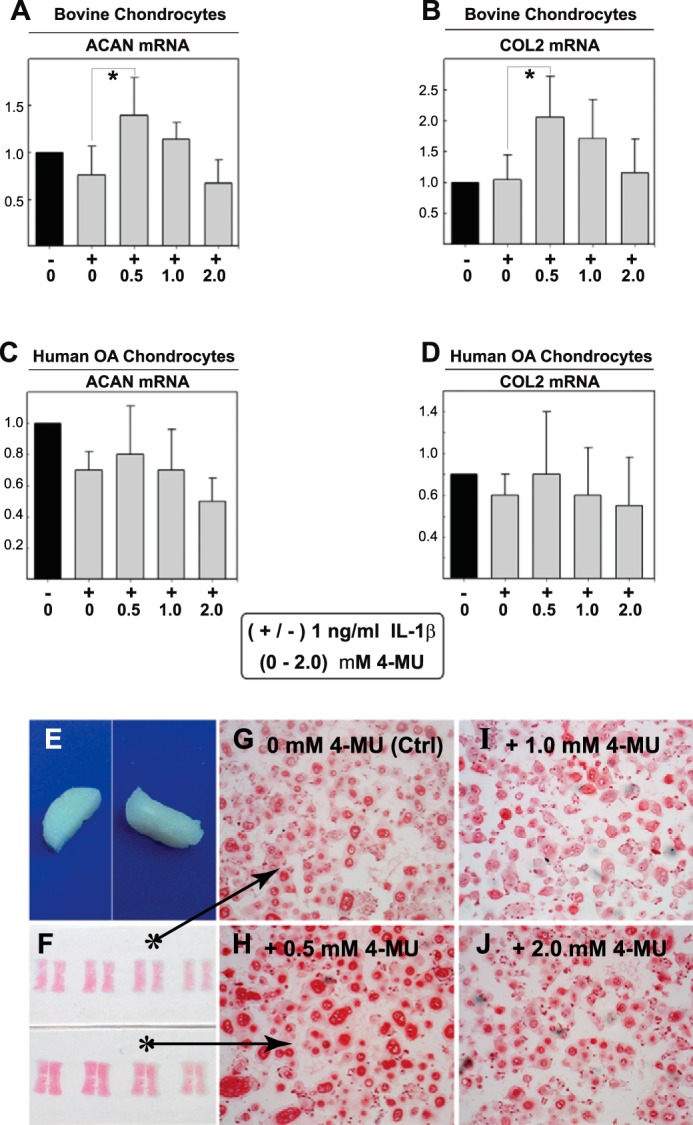
Effects to collagen II and aggrecan of 4-MU treatment of chondrocytes and bioengineered neocartilages. To demonstrate that 4-MU does not exhibit broadly based inhibitory effects on cell metabolism, high density monolayer cultures of bovine chondrocytes (A and B) or human OA chondrocytes (C and D) were treated without or with 1.0 ng/ml IL-1β (−/+) along with co-treatment with 0 to 2.0 mm concentrations of 4-MU as shown. After 24 h, the chondrocytes were lysed and analyzed by real time qRT-PCR. The fold changes (y axes) in mRNA copy number for aggrecan (ACAN, A and C) or collagen II (COL2, B and D) were compared with control (black bars) and quantified using the ΔΔCt approach with normalization to GAPDH. Values represent the average ± S.D. of data derived from three independent experiments. For statistical analysis, a two-way ANOVA followed by Tukey-Kramer test was used; *, p < 0.05; **, p < 0.01. In other experiments, bioengineered cartilage disks were generated from bovine chondrocytes (E) and treated for 72 h without (Ctrl) or with 0.5–2.0 nm 4-MU as labeled (F–J). After treatment, the neocartilage disks were fixed, sectioned, and stained using safranin O/fast green (actual slides shown in F). All images are of equivalent magnification and time exposure settings.