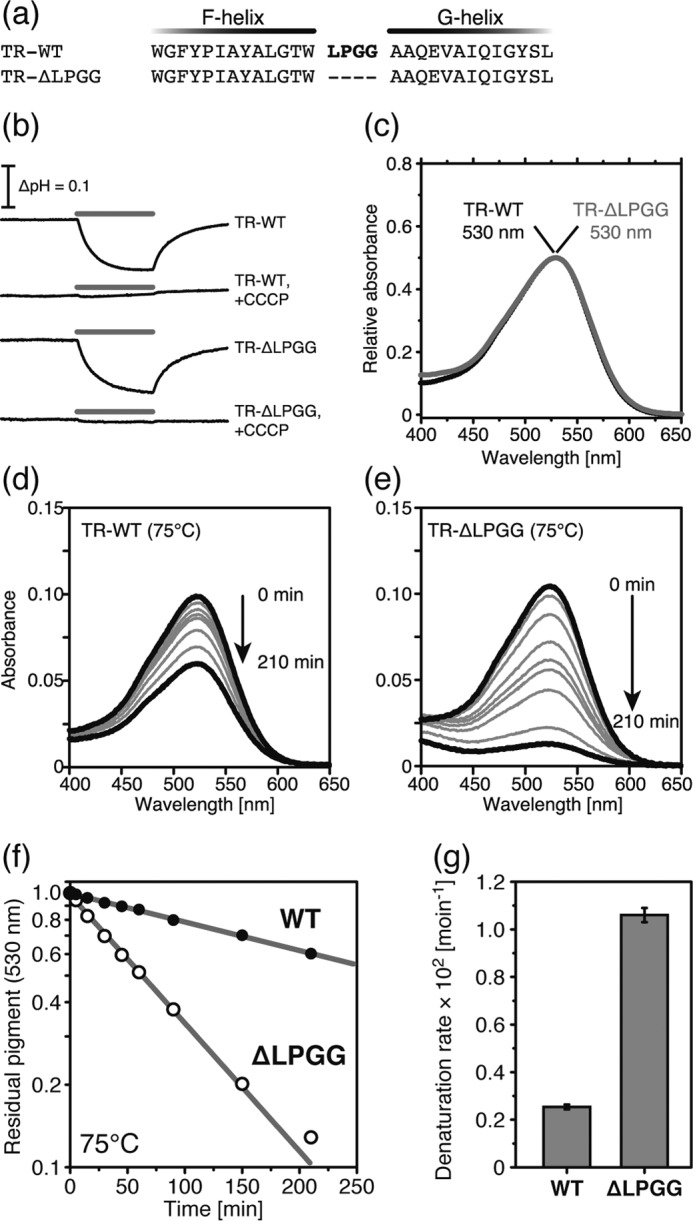
FIGURE 6.

Comparison of TR-WT and TR-ΔLPGG. a, partial amino acid sequence from F- to G-helices. Bold red letters represent the LPGG sequence (Leu211-Pro212-Gly213-Gly21 4) in the extracellular loop between the F- and G-helices. b, light-induced pH changes of TR-WT and TR-ΔLPGG in E. coli suspension in the presence of 100 mm NaCl. The pH was decreased during the 520 ± 10-nm light irradiation for 3 min, shown by the gray bars, in the absence of CCCP. In the presence of 10 μm CCCP, the pH change disappeared. c, visible absorption spectra of TR-WT (black) and TR-ΔLPGG (gray). The absorption maxima were 530 nm. d and e, time-dependent thermal denaturation of TR-WT (d) and TR-ΔLPGG (e) at 75 °C. f, the denaturation kinetics of TR-WT (black) and TR-ΔLPGG (white) at 75 °C. The data were analyzed by a single exponential decay function shown by the gray lines. g, the denaturation rate constants are shown as a bar graph.
