FIGURE 4.
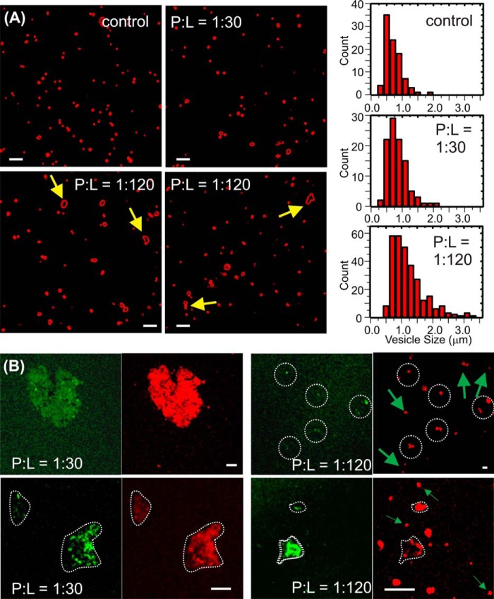
Confocal fluorescence imaging for Aβ-liposome samples. A, representative confocal imaging for rhodamine B-labeled (red channel only) liposomes in the external addition giant unilamellar vesicle samples and the distribution of vesicle sizes after 48-h incubation. The histograms for the control (without peptide) and the sample with 1:30 P:L ratio and 1:120 P:L ratio are shown in the top left, center left, and bottom left panels, respectively. The arrows highlighted non-spherical species that indicate fusion between individual vesicles. B, representative confocal imaging for rhodamine B-labeled (red channel) liposomes and rhodamine green-labeled (green channel) 40-residue Aβ peptides in the preincorporation large unilamellar vesicle samples with a P:L ratio of 1:30 (left panels) and 1:120 (right panels). The white dotted contours highlight the morphologies of aggregates, and the arrows highlight spherical vesicles only composed of lipids (lack of green channel signal at the same locations). Scale bars = 5 μm.

