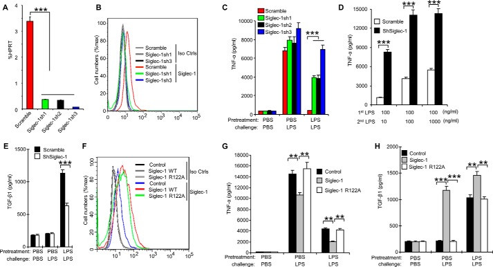
FIGURE 6.
Siglec-1 promotes endotoxin tolerance by enhancing TGF-β1 secretion. RAW 264.7 cells were transduced with lentiviral vector carrying scrambled shRNA or Siglec-1 shRNA. After transduction and selection with puromycin, stable clones expressing the shRNA were isolated and expanded under the selection of 2 μg/ml puromycin. A, real-time PCR analysis of Siglec-1 mRNA level. B, cell surface expression of Siglec-1 on RAW 264.7 was analyzed by flow cytometry. C–E, characterization of RAW 264.7 cells clones stably overexpressing shRNA for Siglec-1. 2 × 10⁵ RAW 264.7 cells were tolerized for 24 h with 100 ng/ml LPS, washed three times with PBS, and then challenged with 1 μg/ml LPS for 6 h (C) or with indicated concentration of LPS for 18 h (D). The culture supernatants were subsequently collected and analyzed for TNF-α (C, D) production as described in Ref. 23–25 and TGF-β1 (E) production by ELISA. F–H, overexpression of Siglec-1 promotes TGF-β1 secretion. F, flow cytometric analysis of Siglec-1 in RAW 264.7 cell clones stably overexpressing Siglec-1. RAW 264.7 cell clones stably overexpressing Siglec-1 were tolerized for 24 h with 100 ng/ml LPS, washed three times with PBS, and then challenged for 16 h with 1 μg/ml LPS. TNF-α in the cell culture supernatants was analyzed with cytokine bead array (G). TGF-β1 in the cell culture supernatants was assessed by ELISA (H). Data presented in this figure have been reproduced at least two times. Data are shown as mean ± S.D.

