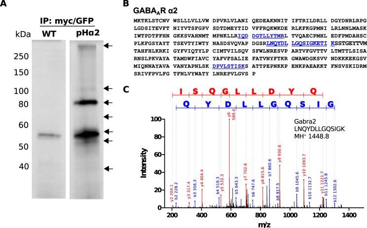
FIGURE 5.
Two-step purification to isolate pHα2 complexes. Detergent-solubilized hippocampal and cortical lysates of age- and sex-matched WT and pHα2 mice were immunoprecipitated with Myc followed by GFP-Trap and subjected to SDS-PAGE and silver staining (A). Representative silver-stained gel depicts bands of interest (arrow) that were excised from pHα2 and the corresponding WT lane for mass spectrometry analysis. Protein coverage of GABAAR α2 subunit (blue, underline) identified by MS analysis (B). Example of MS/MS spectrum for tryptic peptide identified as GABAAR α2 is shown (C). The sequence of the identified peptide is indicated.

