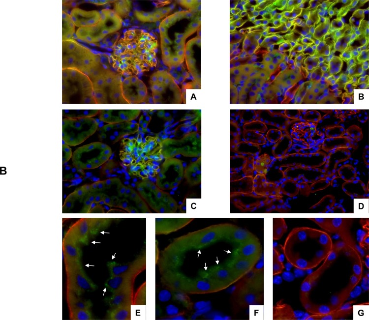
Fig 2. Detection of full length agrin and CAF22 in mouse kidney.
Kidneys from wildtype and NT-/- mice were stained with antibodies against full length agrin (red) and CAF (green). Cell nuclei were marked with DAPI (blue). (A,B,C) Localization of full length agrin and CAF22 in the renal cortex (A,C) and at the transition between outer and inner medulla in wildtype kidney. (D) No staining related to CAF22 was detected in kidneys from NT-/- mice. Original magnification 400x. (E,F) Higher magnification of proximal tubules in wildtype kidney show a punctuate subapical staining for CAF22 (arrows). (G) No subapical staining in proximal tubules of kidneys from NT-/- mice.

