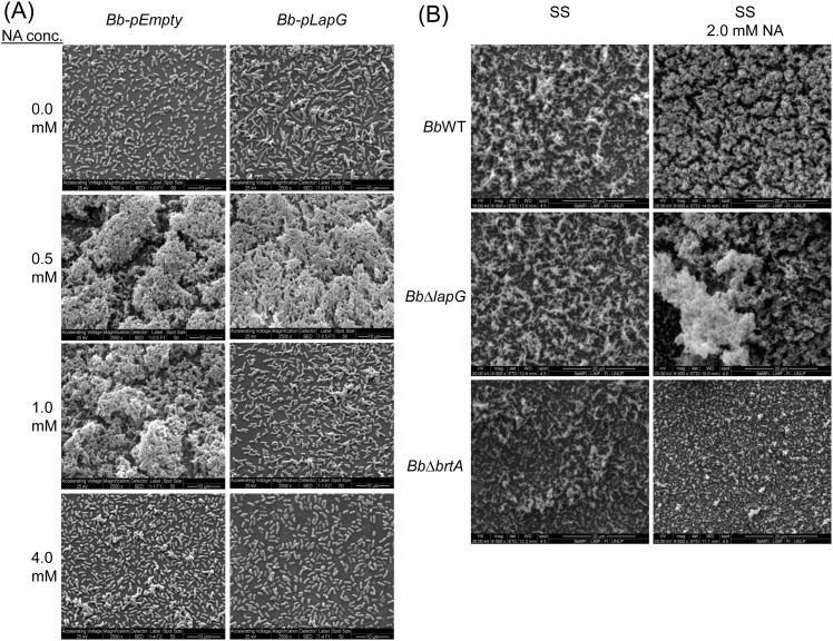
Fig 6. SEM images of B. bronchiseptica biofilms.
(A) The strains Bb-pEmpty (left column of panels) or Bb-pLapG (right column of panels) were grown on vertically submerged coverslips in SS medium alone or supplemented with indicated NA concentrations. After 24 h of growth, biofilms formed at the air–liquid interface were visualized by SEM. (B) Indicated strains were grown on vertically submerged coverslips in SS medium alone (left column of panels) or SS supplemented with 2.0 mM NA (right column of panels).

