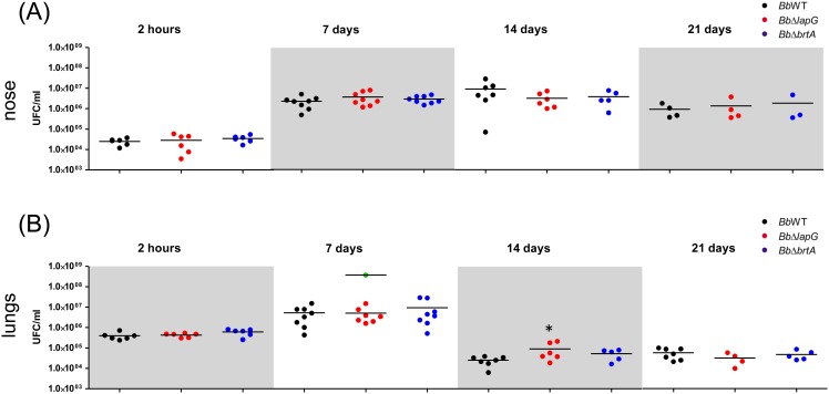
Fig 7. B. bronchiseptica infection in a mouse model.
Comparison of B. bronchiseptica bacterial burden in the mouse nose (A) and lungs (B) were determined as described in the Materials and Methods after the indicated days post-infection. Bacteria were intranasally inoculated into external nares with an air displacement pipette (5 x 10⁵ CFU in 50 μl). Green dots correspond to CFU from euthanized mice infected with BbΔlapG. * indicates a significant difference compared to the CFU determined in mice infected with wild type strain, p<0.01.

