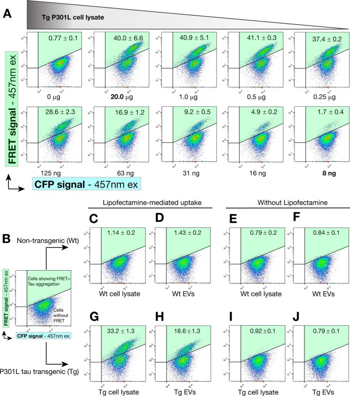
FIGURE 4.
Exosome-like EVs can induce endogenous tau aggregation in FRET tau biosensor cells. FRET tau biosensor cells are HEK293T cells that express both tau-CFP and tau-YFP (RD domain). Cells were treated and analyzed by FRET flow cytometry after a 24-h treatment (n = 3, average ± S.D., 20,000 cells/experiment were analyzed). Representative flow cytometry plots show the FRET signal as detected in the right gate (Q2, shaded in green). A, titration of protein sample to determine concentrations near saturation of FRET tau biosensor cells. Cells were treated with decreasing amounts of rTg4510 transgenic brain cell lysates in the presence of Lipofectamine and analyzed by FRET flow cytometry after a 24-h treatment. 20 μg of protein in cell lysates is sufficient to generate a FRET signal that is not much lower than that obtained with only 1.0 or 0.5 μg of protein. Even with 8 ng of protein, a FRET signal is detected. Therefore, cells are saturated at 20 μg of cell lysate and detect tau seeds up to the low nanogram range. This implies that even minute amounts of tau seeds are detectable in 20 μg of protein. ex, excitation. B–J, analysis of the requirement of Lipofectamine. FRET tau biosensor cells were treated in the presence (C, D, G, and H) or absence (E, F, I, and J) of Lipofectamine, with vehicle (B) showing a lack of signal in the gate for FRET (upper right gate Q2, shaded green). C–F, none of the treatments with WT EVs or cell lysates can induce FRET. G–J, a strong FRET signal is detected with Tg cell lysates (G) and Tg EVs (H), but only in the presence of Lipofectamine. I and J, the same Tg samples without using Lipofectamine.

