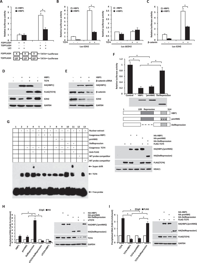
FIGURE 7.
HBP1 decreases EZH2 expression by inhibiting Wnt/β-catenin signaling. A, HBP1 inhibits LiCl-activated gene expression through the LEF/TCF DNA binding site. HEK293T cells transfected with FOPFLASH or TOPFLASH were treated with LiCl for 12 h. Cells were harvested for the luciferase assay. TOPFLASH contains three LEF/TCF sites followed by the minimal TATA and luciferase. FOPFLASH contains three mutated LEF/TCF sites followed by the minimal TATA and luciferase. The luciferase activities were expressed as mean ± S.E. from four experiments. *, p < 0.05. B and C, HBP1 inhibits TCF4 or the β-catenin-activated EZH2 promoter. HEK293T cells were cotransfected with Luc-EZH2 or Luc-ΔEZH2 and TCF4 (B) or the β-catenin (C) expression plasmid. The luciferase activities were expressed as mean ± S.E. from four experiments. *, p < 0.05. D, TCF4 rescues HBP1 inhibition of EZH2 expression. Levels of EZH2 and GAPDH (as a control) were determined by Western blotting in HEK293T cells transfected with HBP1, TCF4, or HBP1 and TCF4. E, β-catenin knockdown by siRNA enhances HBP1 inhibition of EZH2 expression. Levels of β-catenin, EZH2, and GAPDH (as a control) were determined by Western blotting in HEK293T cells transfected with HBP1 with or without β-catenin siRNA. F, repression domain is indispensible for HBP1 inhibition of the EZH2 promoter. HEK293T cells were cotransfected with Luc-EZH2 and wild-type HBP1 or associated mutants. The luciferase activities were expressed as mean ± S.E. from four experiments. *, p < 0.05 (top panel). Anti-HA immunoblots for HBP1 and mutant protein expression are shown (center panel). Also shown is a schematic of wild-type HBP1 and associated mutants (bottom panel). G, HBP1 inhibits TCF4 binding to its affinity site in the EZH2 promoter in vitro. EMSA was performed by using the biotin-labeled probes. Four-microgram amounts of nuclear extracts from HEK293T cells expressing HA-HBP1, HA-pmHMG, or HA-delRepression with or without FLAG-TCF4 were used. The wild-type probe (AGAGTTATATACTGCTTTGATTTGCT) and the mutant probe (AGAGTTATATACTGCGGGTATTTGCT) were used as unlabeled competitors at 100-fold excess. The presence of specific complexes, including supershifted FLAG-TCF4 in the complexes, is indicated (left panel). Anti-HA immunoblots for HBP1 and mutant protein expression and anti-FLAG for TCF4 protein expression are shown (right panel). H, HBP1 binds to the EZH2 promoter through TCF4. H1299 cells were transfected with plasmids indicated in the figure. A ChIP assay was performed with anti-HA antibody or control IgG. The precipitated DNA fragments were amplified with specific oligonucleotides for the EZH2 promoter by real-time PCR. Results are representative of three independent experiments, and values are the mean ± S.E. *, p < 0.05. I, HBP1 inhibits TCF4 binding to its affinity site in the EZH2 promoter in vivo. H1299 cells were transfected with the plasmids indicated in the figure. ChIP assay was performed with anti-FLAG antibody or control IgG. The precipitated DNA fragments were amplified with specific oligonucleotides for the EZH2 promoter by real-time PCR. Results are representative of three independent experiments, and values are the mean ± S.E. *, p < 0.05.

