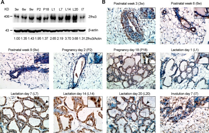
FIGURE 1.
Dynamic expression of Zfhx3 protein at different stages of postnatal mammary gland development. A, detection of Zfhx3 protein by Western blotting in C57BL/6 female mouse mammary tissues at indicated developmental stages. β-Actin served as the loading control. 3w, postnatal week 3; 6w, postnatal week 6; 9w, postnatal week 9; P2, pregnancy day 2; P18, pregnancy day 18; L1, lactation day 1; L7, lactation day 7; L14, lactation day 14; L20, lactation day 20; I7, involution day 7. The data are representative of three mice per group. Band intensities were quantified using the ImageJ program, and ratios of Zfhx3/β-actin, normalized to that of postnatal week 3, are shown at the bottom. B, IHC staining in mammary glands of C57BL/6 female mice at indicated developmental stages. Arrows show Zfhx3-negative body cells in the terminal end bud (postnatal week 3, prepuberty) or Zfhx3-negative ductal luminal cells at postnatal weeks 6 and 9 (puberty) and pregnancy day 2 (early pregnancy); arrowheads show mature alveolar luminal (Zfhx3-positive) cells in pregnancy day 18 (late pregnancy) and lactation days 1, 7, 14, and 20 (lactation). A representative image from three mice is shown for each stage. Scale bar, 50 μm.

