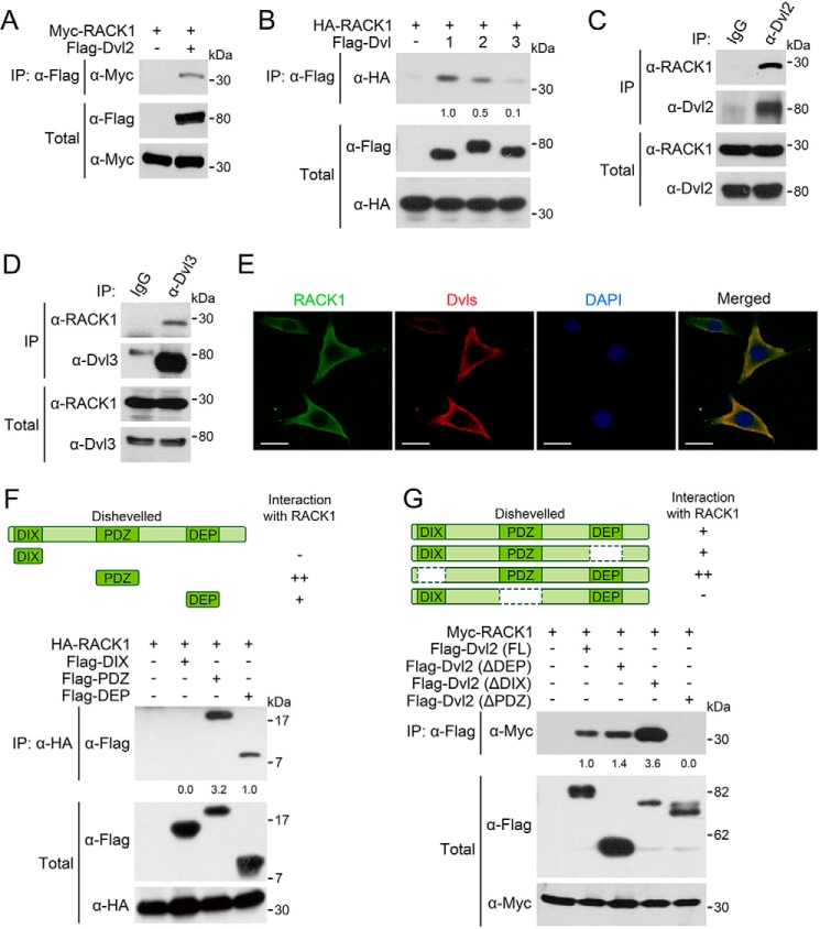
FIGURE 3.
RACK1 interacts with Dvl proteins. A, HEK293T cells were harvested 24 h after being transfected with FLAG-Dvl2 and Myc-RACK1 for anti-FLAG IP followed by IB. The total cell lysates were analyzed by IB with anti-FLAG and anti-Myc antibodies. B, HEK293T cells were harvested 24 h after transfected with FLAG-Dvl1/Dvl2/Dvl3 and HA-RACK1. C and D, HEK293T cells were harvested for IP with anti-Dvl2/Dvl3 antibodies or anti-IgG serum as a control. E, NRK cells were fixed and stained with the indicated antibodies for immunofluorescence. DAPI stained the nucleus. Scale bars = 10 μm. F and G, HEK293T cells were transfected with HA/Myc-RACK1 and truncated Dvl2 fragments. After 24 h, the cells were harvested for anti-HA/FLAG IP followed by IB. The protein levels of precipitated proteins were quantified and normalized against their corresponding total proteins levels, and the values are shown below the bands. All the experiments were independently repeated three times, and a representative one is shown.

