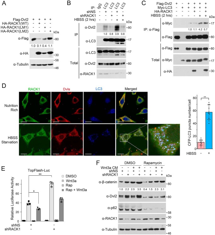
FIGURE 6.
RACK1 enhances the recruitment of Dvl to autophagosome via LC3. A, HEK293T cells were transfected with FLAG-Dvl2 and HA-RACK1 or HA-RACK1 (LM1/2), and cells were then harvested for IB. Tubulin served as a loading control. The protein levels of FLAG-Dvl2 were quantified and normalized against tubulin, and the values are shown below the bands. B, HEK293T cells were infected by a lentivirus expressing nonspecific or RACK1 shRNA. After 48 h, cells were treated with BFA1 (0.2 μm) for 12 h and then treated within HBSS plus BFA1 for another 2 h. Then cell lysates were subjected to IP-IB analysis, and the total cell lysates were analyzed by IB. The protein levels of precipitated Dvl2 were quantified and normalized against precipitated LC3, and the values are shown below the bands. C, HEK293T cells were transfected with FLAG-Dvl2, Myc-LC3, and HA-RACK1 for 24 h and then starved in HBSS for another 2 h. BFA1 (0.2 μm) was added to HBSS for preventing Dvl2 degradation. Then cell lysates were subjected to IP-IB analysis, and the total cell lysates were analyzed by IB. The precipitated Myc-LC3 was quantified and normalized against precipitated FLAG-Dvl2, and the values are shown below the bands. D, NRK cells stably expressing CFP-LC3 were treated with HBSS containing 0.2 μm BFA1 for 2 h and then fixed and stained with anti-Dvl and anti-RACK1 antibodies for immunofluorescence. The CFP-LC3 puncta in each cell (n = 10 cells from three different slides) were counted, and the number of puncta per cell is represented as the mean ± S.D. in the right panel. Two-tailed unpaired Student's t test was used in statistical analyses (**, p < 0.01). Scale bars = 10 μm. E, the experiment was conducted similarly as in Fig. 1D, and rapamycin (Rap, 2 μm) was added to induce autophagy. Luc, luciferase. F, HEK293T cells were infected by a lentivirus expressing nonspecific or RACK1 shRNA. After 48 h, the cells were treated with Wnt3a CM and/or rapamycin (2 μm) for 6 h and then harvested for IB. p62 was used as an autophagy marker. The protein levels of β-catenin were quantified and normalized to tubulin, and the values are shown below the bands.

