Fig. 2.
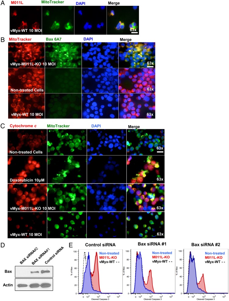
M011L protein localizes to mitochondria in brain-tumor initiating cells (BTICs) infected with vMyx-WT and prevents cytochrome c release by associating with proapoptotic protein Bax. (A) BTIC25 cells were infected with vMyx-WT (10 multiplicity of infection [MOI] for 24 hours prior to co-staining for M011L, mitochondria (Mitotracker Green FM) and nuclei (DAPI). Cells were visualized using an automated Zeiss Observer Z.1 inverted microscope through a 63X/1.4NA objective. M011L protein shows mitochondrial localization in cells infected with vMyx-WT. The data are representative of 3 independent experiments. (B) BTIC48 cells were infected with vMyx-WT (10 MOI), vMyx-M011L-KO (10 MOI), or left untreated for 6 hours prior to staining with an Alexa Fluor 488-labeled antibody to Bax (clone 6A7), Mitotracker Red, and DAPI. Note the co-localization (yellow) of active Bax with the mitochondria following infection with vMyx-M011L-KO (row 1, panel 4). Data represent 3 independent experiments. (C) BTIC48 were infected with vMyx-WT (10 MOI), vMyx-M011L-KO (10 MOI), treated with doxorubicin (10 μM) or left untreated for 10 hours prior to co-staining for cytochrome c (Alexa Fluor 546, red), mitochondria (MitoTracker Green FM), and nuclei (DAPI). Note increased staining for cytochrome c in cytosol (yellow) following treatment with doxorubicin (row 2, panel 4) and vMyx-M011L-KO (row 3, panel 4). Scale bar 25 µm. (D) BTIC48 cells were transfected with siRNAs specific for Bax or a control nontargeting pooled siRNA. After 48 hours, cells were collected, and Bax expression was determined by immunoblotting. (E) In parallel, 48 hours after transfection with siRNAs, BTICs were infected for 72 hours with either vMyx-M011L-KO (10 MOI), vMyx-WT (10 MOI), or left uninfected prior to assessing caspase 3 activation by flow cytometry.

