Fig. 3.
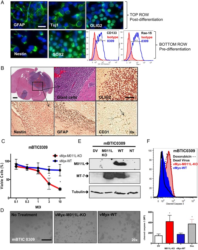
Development and in vitro characterization of murine BTIC0309. (A) In vitro, neurospheres express neuronal markers SOX2, nestin, CD133 (Prominin-1) and Rae-1δ (NK-cell ligand, lower right fluorescence-activated cell sorting [FACS]) and can be differentiated in serum into glial (GFAP), neuronal (Tuj1), and oligodendrial (OLIG2) cell types. Scale bar 25 µm. (B) Mouse brain cross-section after intracranial implantation of mBTIC0309 at day 35 post infection. Scale bar 2 mm. Higher magnification of tumor tissue depicts cytonuclear pleomorphism and giant cells (upper middle). Tumors express endothelial cell marker CD31 and neuronal markers and maintain stem cell characteristics, as seen by immunohistochemistry of OLIG2, nestin and GFAP (brown staining) Scale bar 200 µm. (C) Alamar Blue assay of mBTIC0309 72 hours after infection with vMyx-WT or vMyx-M011L-KO. Data are mean ± SEM of experiments performed in triplicate, and results are representative of at least 3 independent experiments (*; P< .05 by Student t test). (D) vMyx-M011L-KO-treated mBTIC0309 shows substantial evidence of cytopathic effects compared with vMyx-WT treated cells. Neurospheres were imaged using a Zeiss Axiovert microscope at 20× magnification, 72 hours post infection. Data represent 3 independent experiments. (E) Immunoblot 24 hours after vMyx-WT or vMyxV-M011L-KO infection for M011L, MT-7 (early viral gene expression), and tubulin (protein loading control). (F) mBTIC0309 were infected with vMyx-M011L-KO (10 MOI), vMyx-WT (10 MOI), or UV-inactivated (dead) virus or treated with doxorubicin (10 μM). Cells were collected after 72 hours, fixed, and stained with an antibody for cleaved caspase 3 and analyzed by flow cytometry. Data are representative of 3 independent experiments. Means ± SD from triplicates are shown (*P< .05 by ANOVA compared with dead virus).

