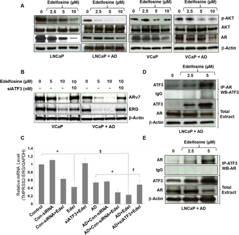
Figure 3. Changes in AKT, AR, ARv7 and ERG protein levels after AD and edelfosine treatments.
(A) LNCaP and VCaP cells were cultured in complete or AD medium for 3 days in 10 cm tissue culture dishes and then treated with edelfosine (0, 2.5, 5 and 10 μM). Treated cells were harvested after 24 h, whole-cell lysates were extracted, and the expression of p-AKT, total AKT and AR were determined by Western blotting. The same blot was probed again for β-actin. (B) VCaP cells grown in complete or AD medium were pre-transfected with siATF3 (10 nM) for 18 h in the presence of lipofectin reagent followed by edelfosine (0, 5 and 10 μM) treatment. Whole-cell lysates were extracted from treated cells after 24 h. ARv7 and ERG expression was determined by Western blotting and the same blot was stripped and re-probed for β-actin. (C) Relative mRNA levels of TMPRSS2-ERG gene product ERG in VCaP cells. Real-time PCR was performed using total RNA extracted from VCaP cells. The cells grown in CM or AD media were treated with edelfosine (10 μM), siATF3 or control siRNA (10 nM) either alone or in combination with edelfosine. The results were normalized to the expression of GAPDH and presented as relative mRNA expression levels (mean ± SD, n = 3). *p < 0.001, compared to respective controls, ‡p < 0.05, compared to edelfosine alone, †p < 0.05, compared to AD + edelfosine (one-way ANOVA). (D) ATF3 interaction with AR. AD and edelfosine (0, 2.5 and 5 μM) treated whole-cell lysates were co-immunoprecipitated with antibodies against AR, followed by Western blot analysis with antibodies specific for ATF3. (E) Reverse immunoprecipitation was carried out in the AD and edelfosine (0, 2.5 and 5 μM) treated whole-cell lysates to confirm the interaction between ATF3 and AR.

