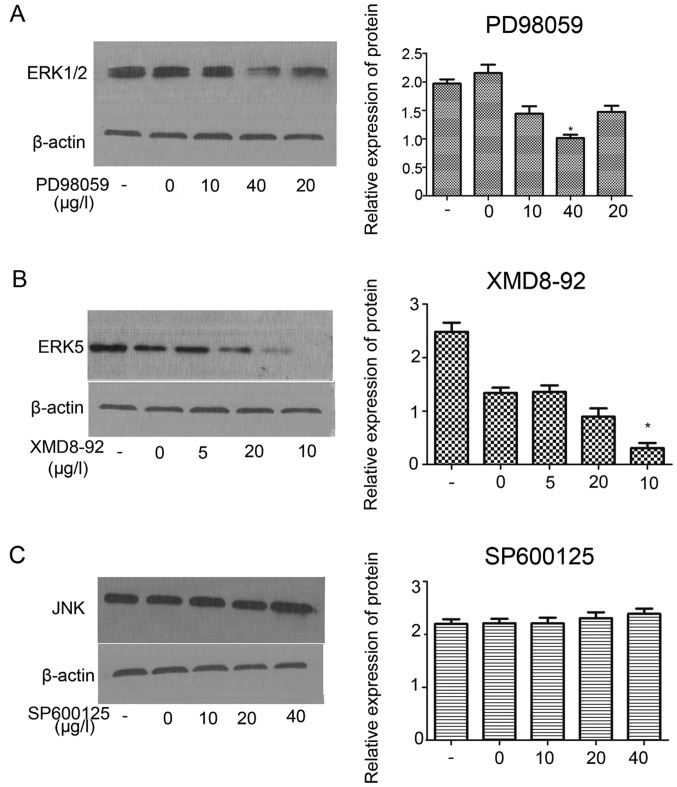
Figure 7.
Inhibitions of protein expression of ERK1/2 and ERK5 through ERK1/2 (MAPK) pathways in HER-2-positive IOMM-Lee meningioma cells. Protein level of ERK1/2 in the HER-2-over IOMM-Lee cells were determined using western blot analysis at 72 h post-transfection. β-actin was used as an internal loading control. (A) Western blot assay demonstrated that PD98059 (ERK1/2 inhibition) inhibited the protein expression of ERK1/2 at the best inhibition concentration of 40 µg/l (*P<0.05). (B) Western blot assay demonstrated XMD8-92 (ERK5 inhibition) inhibited the protein expression of ERK5 the component of ERK1/2 signaling at the best inhibition concentration of 10 µg/l (*P<0.01). (C) No effect we can observed on the JNK at different inhibition concentration of SP600125. The data are expressed as the mean standard deviation from three independent experiments. -, Blank group; 0, the inhibition of 0 µg/l; 5, the inhibition of 5 µg/l; 10, the inhibition of 10 µg/l; 20, the inhibition of 20 µg/l; 40, the inhibition of 40 µg/l.

