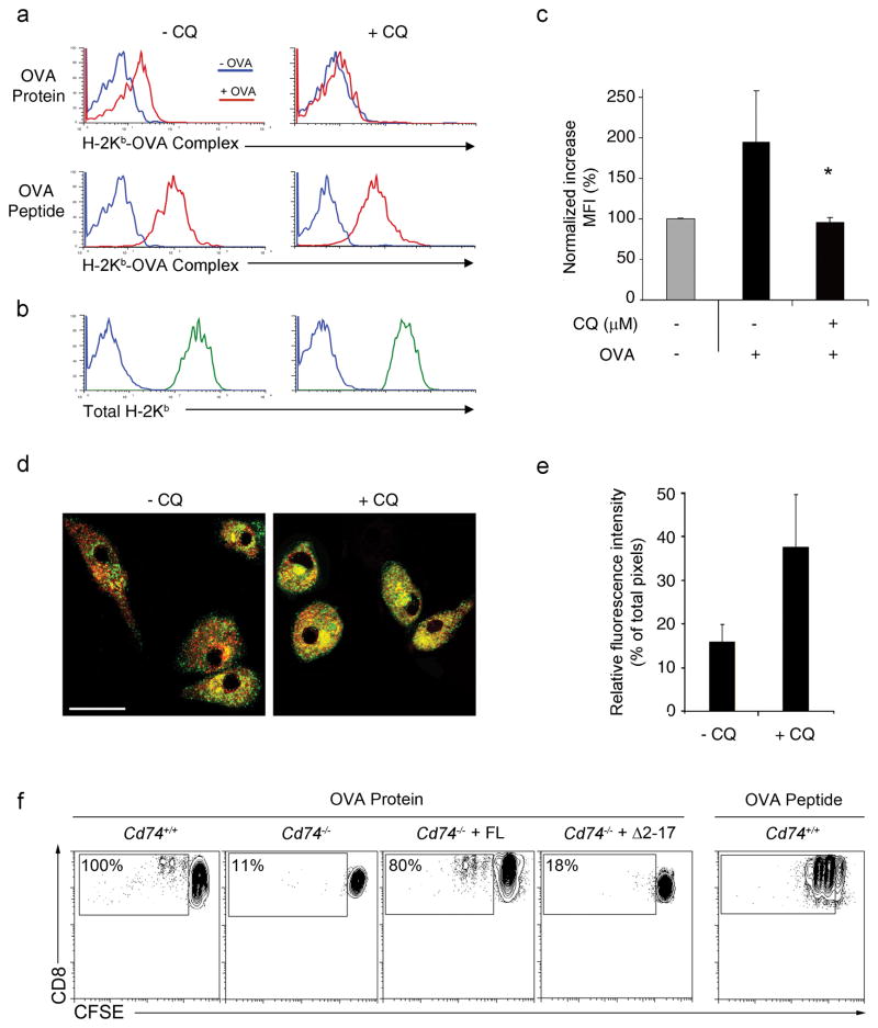
Figure 5. Inhibition of CD74-mediated MHC I trafficking in DCs by treatment with chloroquine.
(a) BMDCs were treated with CQ and the formation of H-2Kb-OVA257-264 complexes on DCs following incubation with soluble OVA (red; top panel) or OVA peptide (red; bottom panel) compared to media alone (blue) was measured by flow cytometry. (b) Total H-2Kb (green) above background (blue) was also assessed. (c) The fold increase in surface H-2Kb-OVA257-264 complexes following incubation with soluble OVA was quantified. Graphs depict normalized increases in mean fluorescence intensity (MFI) ± SD. * p < 0.05. (d) Mature BMDCs treated with CQ were costained with H-2Kb (red) and CD74 (green) antibody. The figure shows optically merged images representative of the majority of cells examined by ICM. Scale bar, 5 μm. (e) Quantitative assessment comparing H-2Kb colocalization with CD74. Graph depicts normalized individual color pixel percentages divided per total pixels ± SD. (f) Cd74−/− BMDCs reconstituted with full length (FL) CD74 or truncated (Δ2–17) CD74 lacking the endolysosomal trafficking motif were incubated with soluble OVA and assessed for the ability to induce proliferation in CFSE-labeled OT-I cells. Percentages represent the proportion of proliferating OT-I cells normalized to Cd74+/+ controls.

