
Figure 1.
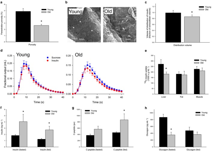
(a) Decreased porosity of the LSEC with age from scanning electron micrographs (n = 5 young and 5 old F344 rats P = 0.006). (b) Sample scanning electron micrographs of the LSEC to clearly illustrate the difference between young and defenestrated old rats, original micrographs taken at 15 000× magnification (fenestrations are indicated by an *). (c) There is a 20% reduction in the fractional volume of distribution of insulin with age (n = 9 young and 10 old F344 rats, P = 0.01) (d) MID outflow curves for insulin and the extracellular marker sucrose. Insulin exits the liver prior to sucrose in the old animals, indicating a restricted access to the entire extracellular space with age‐related defenestration. (e) 14C‐insulin uptake by the liver was found to be significantly reduced with age, but was found to be unchanged in the muscle and the fat (n = 10 young and 10 old mice, P < 0.05). (f) Fasting and fed insulin levels were found to be significantly elevated with age in C57Bl6 mice (n = 6 young and 5 old mice, P < 0.05). (g) C‐peptide levels were found to be significantly elevated with age in the fed state (n = 4 young and 5 old mice, P = 0.02) and (h) Glucagon levels were found to be suppressed in the fasting state in old mice (n = 6 young and 6 old mice, P = 0.02).