Figure 1.

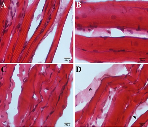
Compound panel of hematoxylin-eosin images of contralateral (A, B) and ipsilateral (C, D) masseter muscles. A) Low magnification of contralateral muscle showing normal and rectilinear fibers morphology. B) High magnification of contralateral muscle showing high number of nuclei within the fibers and along sarcolemma. C) Low magnification of ispilateral muscle showing non-healthy convolute fibers morphology, some atrophic fibers (arrowhead). D) Image of ipsilateral muscle showing the loss of contractile elements within the fiber (asterisk).
