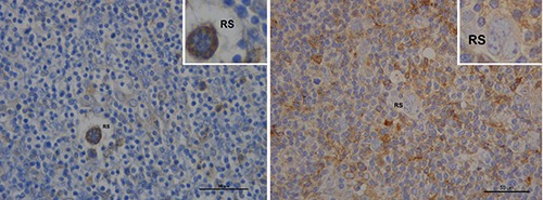
Figure 2.

HLA-G expression (brown color) according to PET-2 results. Left: a 45 year-old female with negative PET-2. The patient shows low HLA-G protein expression in lymphocytes; conversely, Reed-Srernberg cells (RS) are stained. Right: HLA-G is more expressed in reactive cells then in Reed-Srernberg cells (RS) in a 37 year-old female with positive PET 2. Scale bars: 50 μm.
