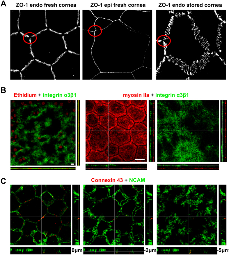
Figure 2. Additional features of human corneal endothelial cells (CECs).
(A) Specificity of ZO-1 expression in CECs in fresh and stored corneas compared to superficial epithelial cells. The zig-zag pattern observed in a fresh cornea (65 years old patient with stromal dystrophy) was dramatically amplified in a cornea placed for 35 days in organ culture medium (74 years old donor with normal cornea). In both cases, ZO-1 remained absent at the Y junctions between cells (red circles), in contrast with the uniform distribution in epithelial cells. (B) Integrin α3β1 was almost evenly expressed in the basal membrane, following the slight ripples of the underlying Descemet’s membrane. Both samples were from 21 and 27 year-old patients, grafted for keratoconus. Scale bars = 10 μm. (C) Labeling of gap junctions (connexin 43) showed discontinuous points situated mainly in the subapical lateral membranes (revealed by NCAM); it could be found at intermediate levels but seldom at basal levels.

