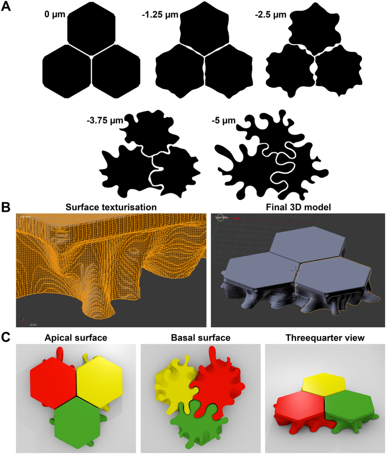
Figure 5. 3D reconstruction of human corneal endothelial cells (CECs).
(A) Five layers uniformly distributed over the CEC height were schematized using information provided by confocal images of membrane proteins. (B) Example of surface smoothing using the freeware blender and final 3D reconstruction of 3 neighbor CECs. (C) Different view of 3D printed cells for pedagogic purpose. The simplified model is consistent with the characteristics of cell-cell junctions: straight borders with holes at the Y junctions at the apical surface, interconnected intercellular spaces between cells, tightly joined interdigitating foot processes at the basal surface. Only the number of membrane folds and interdigitations might have been underestimated (see N-cadherin Fig. 2B).

