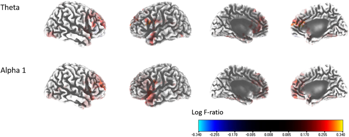
Figure 1. Source-localized correlation analysis between the percentage improvements in the Tinnitus Handicap Index (THI) score and the pre-tinnitus retraining treatment (TRT) resting-state quantitative electroencephalography data.
The percentage improvements in the THI score were positively correlated with the activities of the left medial frontal cortex [Brodmann area (BA) 9], the left rostral anterior cingulate cortex (rACC; BA 24), and the right dorsolateral prefrontal cortex (DLPFC; BA 10) (i.e., the theta frequency band); and the activities of the the left insula (BA 13), the right DLPFC, the left rACC, the left pregenual anterior cingulate cortex (BA 32), and the left inferior frontal gyrus (BAs 45 and 47) (i.e., the alpha 1 frequency bands).

